
Optical Coherence Tomography (OCT)
OCT is a detailed, painless scan of the retina's individual layers - and at The Eye Clinic at Rondels, it's included as standard with every eye examination, not an optional extra.
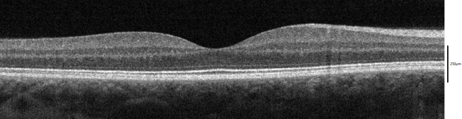
A real OCT cross-section of a healthy macula, showing the foveal dip and distinct retinal layers. Image: Wies6014, Wikimedia Commons, adapted (cropped), licensed under CC BY-SA 4.0.
What is OCT?
Light, not sound
OCT works like an ultrasound scan, but uses light instead of sound waves - producing a far more detailed, microscope-level cross-section of the retina rather than a blurry outline.
Every layer of the retina
The retina is made up of multiple distinct layers, each doing a different job - OCT images them individually, to a precision of a few thousandths of a millimetre.
Seconds, not minutes
The scan itself is painless, doesn't touch the eye, and takes just a few seconds per eye - comfortably part of a normal appointment, not a separate visit.
Included as standard, every time
Many practices only offer OCT as a paid add-on. We include it as a routine part of every eye examination here, because it catches so much that a standard examination alone simply can’t - without ever adding to your appointment length in any meaningful way.
What OCT can reveal, often years early
Many of these conditions have no symptoms at all in their early stages - OCT is often the only way to catch them this soon.
Glaucoma
Measures the thickness of the nerve fibre layer around the optic nerve - often showing early glaucoma damage before it would ever affect your visual field test.
Macular disease
Age-related macular degeneration, macular holes and epiretinal membranes all show up clearly as changes to the retina's normal layered structure.
Diabetic eye disease
Detects the earliest signs of fluid and swelling in the retina from diabetic maculopathy, often well before it would be visible on a standard retinal photograph.
Retinal detachment risk
Can reveal early retinal thinning, holes or traction that may otherwise go unnoticed, so any risk can be monitored or referred on promptly.
See our Eye Health Conditions and Adult Eyecare pages for more on glaucoma and macular disease specifically.
Part of every eye examination
You don’t need to ask for an OCT scan or book it separately - it’s simply part of what a thorough eye examination looks like here.
Book an eye examination